
Page 166 - Contributed Paper Session (CPS) - Volume 2
P. 166
CPS1494 Senthilvel V. et al.
known Type II DM patients and to find the associated risk factors of DR among
known Type II DM patients.
2. Materials and Methods
We have conducted a hospital-based cross-sectional study among known
Type II DM patients on DR in the Department of Endocrinology, Amrita
Institute of Medical Sciences and Research Institute, Kochi, Kerala from
nd
1 February to 2 March, 2018 with a sample size of 150 Type II DM
st
patients. Patient’s selection for the study: 350 patients were attended and
took treatment in the Department of Endocrinology. Patients those who were
under treatment for DM was consecutively selected by an ophthalmologist
were included in our present study. An inclusion criteria of the patients with
aged 30 years and above those who were having DM for at least 1 year and
above and who are all the residence of Kochi area, Kerala, South India and with
an exclusion criteria the patients those who were having chronic diseases.
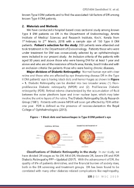
Major divisions of Diabetic Retinopathy: Normal human eye with clear
retina and those who are affected by eye threatening disease DR in the Type
II DM patients’ eye is having black dots and hemorrhages as shown in Figure
– 1. Diabetic Retinopathy can be divided into two broader areas: (1). Non-
proliferative Diabetic retinopathy (NPDR) and (2). Proliferative Diabetic
retinopathy (PDR). Retinal edema characterized by the accumulation of fluid
between the outer plexiform layer and inner nuclear layer, which may later
involve the entire layers of the retina, The Diabetic Retinopathy Study Research
Group (1981). Patients with severe NPDR will soon get affected by PDR within
one year. PDR is defined as the presence of neovascularization the Royal
College of Ophthalmologists (2013).
Figure – 1 Black dots and hemorrhages in Type II DM patient’s eye
Classifications of Diabetic Retinopathy in the study: In our study, we
have divided DR stages as No DR, Mild DR, Moderate DR, Severe DR and PDR
Diabetic Retinopathy PPP – Updated (2017). With the advancement of DR, the
quality of life of patients diminishes, and the financial burden of society rises,
both in the DR screening and treatment groups. DR has been found to be
correlated with many other diabetes-related complications like nephropathy,
155 | I S I W S C 2 0 1 9